 If you’ve received a skin cancer diagnosis, Mohs surgery is likely to be the preferred method of treatment. Mohs surgery is efficient, precise, and has up to a 99% cure rate for a skin cancer that has not been treated before, and up to a 94% cure rate for a skin cancer that has recurred after previous treatment.*
Mohs surgery is often considered the most effective technique for most types of skin cancers, including melanoma, because it involves minimal discomfort, and encourages the greatest preservation of healthy tissue, which means less risk of scarring and superior cosmetic results.
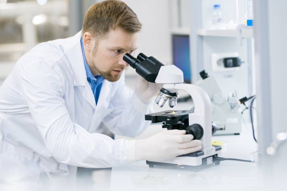
Mohs surgery is conducted in stages by a licensed and specially trained dermatologist or surgeon. During the procedure, the cancerous tissues are removed layer by layer. After removing a layer of tissue, the surgeon examines it under a microscope in an on-site lab. If any cancer cells remain, the surgeon removes another layer of tissue. This is repeated until a layer shows no signs of cancer and all the cancer is removed.
The biggest difference between Mohs surgery and routine excisional surgery is that Mohs is done in stages, and lab results are immediately available to the surgeon on-site, rather than sending a tissue sample to a lab for results that wouldn’t likely be available for days to weeks.
As incidences of skin cancer continue to rise, it’s important to be well-informed and ready to make the best decision for your skin in your time of need. If left untreated, skin cancer can progress to the point of being life-threatening.
Trust Florida Dermatology and Skin Cancer Centers with the diagnosis and treatment of skin cancer with Mohs surgery. Call us today for more information and to schedule a comprehensive skin exam.
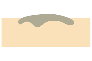 Step 1 When skin cancer is diagnosed, we know the tumor’s roots are deeper than we can see. We have to remove the roots to prevent a recurrence.
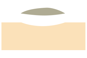 Step 2 The section of tumor we can see is surgically removed.
 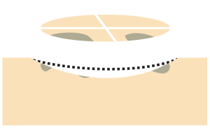 Step 3 A layer of skin is removed. The surgeon divides the tissue into segments, color-codes them with dye, and marks the skin to show where each section came from. The surgeon then draws a map of the surgical site.
Step 4 A layer of skin is removed. The surgeon divides the tissue into segments, color-codes them with dye, and marks the skin to show where each section came from. The surgeon then draws a map of the surgical site.  Step 5 If cancer cells are identified with the microscope, the surgeon reviews the map and removes another layer of skin, but only from the precise place the remaining cancer cells were found.
 Step 6 The procedure continues in this manner until all evidence of cancer is gone. By removing only tissue that contains cancer, the healthy skin remains intact and less scarring occurs.