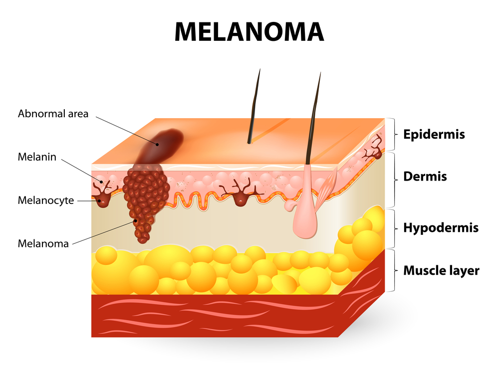
Melanoma is a type of skin cancer that forms when cells in the upper layer of skin called melanocytes start to make some abnormal changes. Melanocytes produce the pigment that gives our skin its color. This pigment is called melanin, and it also is responsible for helping to protect us from harmful exposure to ultraviolet (UV) rays. When cellular DNA modifications take place and melanocytes start to grow in an anomalous fashion, melanomas can begin to form.
Melanoma is the most dangerous and aggressive form of skin cancer. Left untreated, it can spread quickly and become fatal. To detect melanomas, follow the ABCDE rule and look for growths that are:
- Asymmetrical in size
- Borders are irregular or jagged
- Color is not the same all over, and includes areas of blue, brown, black, or red
- Diameter is larger than the size of a pencil eraser (6 mm)
- Evolving in size, shape, or color
Early identification and diagnosis of cancerous lesions increases the likelihood that treatment will be successful. To make a diagnosis, your dermatologist will perform a skin examination, and determine next steps.
Depending on the size, location and severity of the growth, several treatment options are available to remove cancerous cells, including Mohs micrographic surgery, the most effective technique for most types of skin cancers, with minimal scarring or risk.
FLDSCC brings an unmatched level of services to all our patients, and we are confident in our approach to treat skin cancer most effectively and efficiently. Fortunately, skin cancer treatment, specifically melanoma, has a very favorable outcome since if it is detected early, it’s almost always treatable.
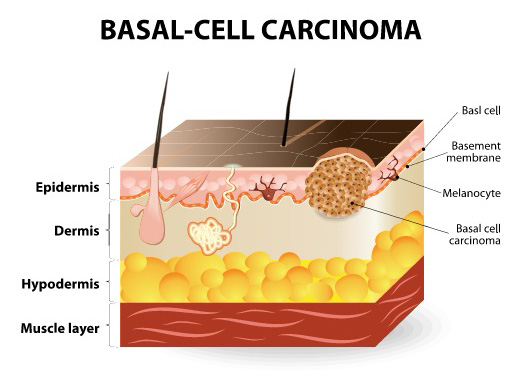
Basal cell carcinoma (BCC) is the most common form of skin cancer, and is known to frequently recur. BCC cells are slow to grow and have a high curability rate. Typically these cancerous cells are found on areas of the skin that have been exposed to the sun’s harmful ultraviolet rays. BCCs can vary in appearance from person to person.

BCCs can appear as one or more of the following:
- Red or pink patches or lumps
- Open sores that never heal
- A scar-like area of the skin that appears white or yellow
- Shiny bumps
- Elevated growths
- Growths with an indent
- Brownish scars or flesh-colored lesions
If left untreated, skin cancer can progress to the point that it is disfiguring and even life threatening. Early detection and treatment of skin cancers saves lives.
For more information about how to treat basal cell carcinoma and to schedule an annual skin exam, contact us today.
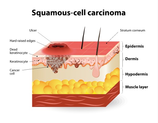
Squamous cell carcinoma (SCC) is the second most common type of skin cancer, and very treatable if identified and diagnosed early. Squamous cell carcinomas are more likely than basal cell carcinomas to spread to other parts of the body. They can also grow deep, causing bone and nerve damage.
The most frequent cause of SCC is exposure to ultraviolet radiation (UV) from the sun and tanning beds. SCCs often develop in areas of skin injury or damage – like scars, sores, age spots, and wrinkles.
SCCs may present as:
- A firm red bump
- A flat, rough, scaly lesion – bleeding and crusting may be present
- A sore that doesn’t heal, or heals and reopens
Squamous cell carcinomas can develop from precancerous lesions called actinic keratosis (AK). AKs are rough, scaly patches that develop on sun-exposed skin after years of sun exposure. AKs have a high risk of progressing into skin cancer – commonly squamous cell carcinoma.
Early detection is crucial for successful treatment of skin cancer. For more information about services that Florida Dermatology and Skin Cancer Centers provides, or to make an appointment for a skin exam, contact us today.


